Home

kalligraf klon Mission skin histology h&e fjols der ovre Forretningsmand

Human Plantar Skin Slide, 7 m, H&E: Amazon.com: Industrial & Scientific

H E Stain: Most Up-to-Date Encyclopedia, News & Reviews

Histology of skin cross sections via H&E staining 24 hours after DNA... |  Download Scientific Diagram
Histology of skin cross sections via H&E staining 24 hours after DNA... | Download Scientific Diagram

Histomorphology and Vascular Lesions in Dorsal Rat Skin Used as Injection  Sites for a Subcutaneous Toxicity Study - Monique Y. Wells, Hélène Voute,  Valérie Bellingard, Cécile Fisch, Virginie Boulifard, Catherine George,  Philippe
Histomorphology and Vascular Lesions in Dorsal Rat Skin Used as Injection Sites for a Subcutaneous Toxicity Study - Monique Y. Wells, Hélène Voute, Valérie Bellingard, Cécile Fisch, Virginie Boulifard, Catherine George, Philippe

H&E staining of human skin. This photograph of H&E-stained tissue... |  Download Scientific Diagram
H&E staining of human skin. This photograph of H&E-stained tissue... | Download Scientific Diagram

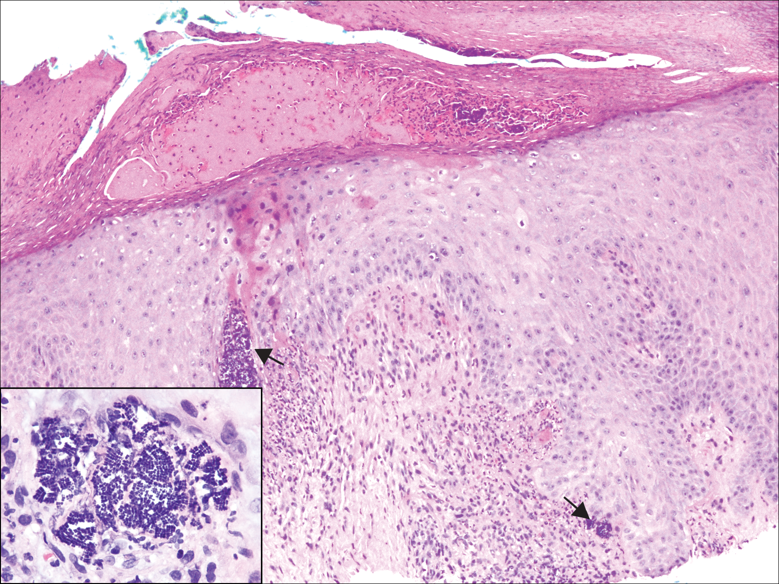
Tetrad Bodies in Skin | MDedge Dermatology

A novel method for tissue segmentation in high-resolution H&E-stained  histopathological whole-slide images - ScienceDirect
A novel method for tissue segmentation in high-resolution H&E-stained histopathological whole-slide images - ScienceDirect

Haematoxylin and Eosin (H and E) stain of normal skin as control. There...  | Download Scientific Diagram
Haematoxylin and Eosin (H and E) stain of normal skin as control. There... | Download Scientific Diagram

H&E stain - Wikipedia

A novel method for tissue segmentation in high-resolution H&E-stained  histopathological whole-slide images - ScienceDirect
A novel method for tissue segmentation in high-resolution H&E-stained histopathological whole-slide images - ScienceDirect
Blue Histology - Integumentary System

Skin: The Histology Guide

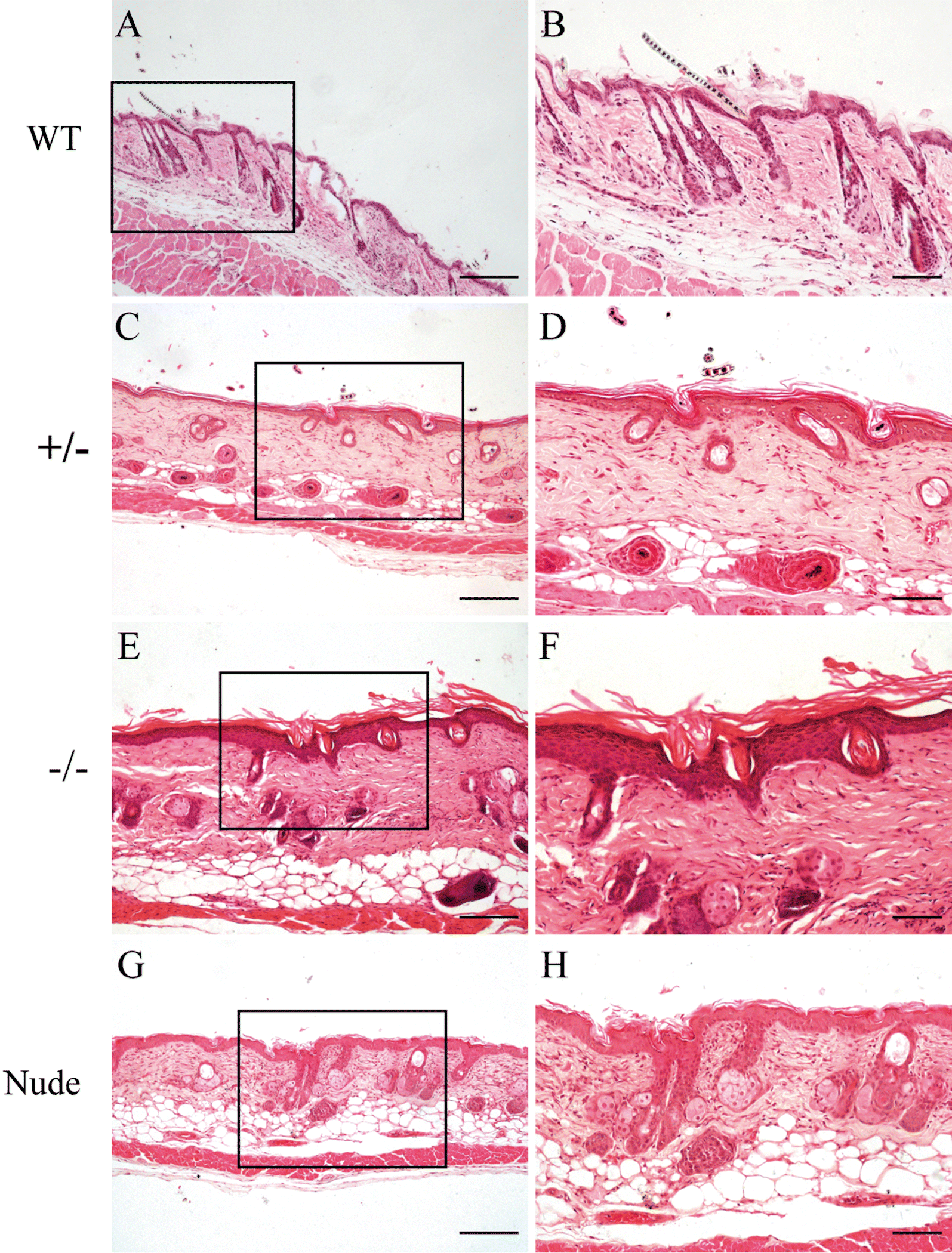
Abnormalities of hair structure and skin histology derived from  CRISPR/Cas9-based knockout of phospholipase C-delta 1 in mice | Journal of  Translational Medicine | Full Text
Abnormalities of hair structure and skin histology derived from CRISPR/Cas9-based knockout of phospholipase C-delta 1 in mice | Journal of Translational Medicine | Full Text

Histology of Skin Stained with H&E Stock Photo - Image of skin,  hematoxylin: 42769166
Histology of Skin Stained with H&E Stock Photo - Image of skin, hematoxylin: 42769166

Human Heavily Pigmented Skin sec. 7 m H&E Stain Microscope Slide:  Amazon.com: Industrial & Scientific
Human Heavily Pigmented Skin sec. 7 m H&E Stain Microscope Slide: Amazon.com: Industrial & Scientific
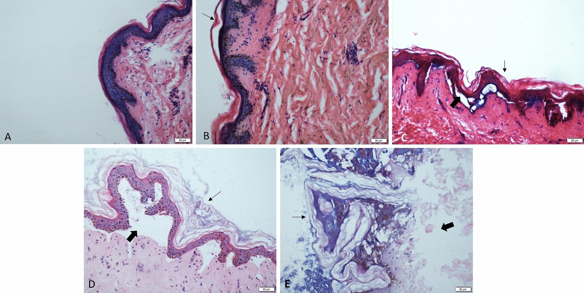
HistoQuarterly: SKIN | Histology Blog

Tip Chapter: Histology and Physiology of the Skin | SpringerLink

Histological changes in human skin 32 days after death and the potential  forensic significance | Scientific Reports
Histological changes in human skin 32 days after death and the potential forensic significance | Scientific Reports

Normal skin histology. Histology shows epidermis (E), dermis (D) and... |  Download Scientific Diagram
Normal skin histology. Histology shows epidermis (E), dermis (D) and... | Download Scientific Diagram

Hairy skin, longitudinal section (H&E Staining) - PUMS Histology Slides  Review Series - YouTube
Hairy skin, longitudinal section (H&E Staining) - PUMS Histology Slides Review Series - YouTube

Hematoxylin & eosin (H&E) staining of micropig skin. The histology is... |  Download Scientific Diagram
Hematoxylin & eosin (H&E) staining of micropig skin. The histology is... | Download Scientific Diagram

Skin histology. H&E-stained longitudinal sections of tail ( A – I ) and...  | Download Scientific Diagram
Skin histology. H&E-stained longitudinal sections of tail ( A – I ) and... | Download Scientific Diagram

Histological and immunohistochemical examination of mouse skin treated with  and without imiquimod cream: (A) hemoxylin and eosin (H&E) staining; (B)  the epidermal thickness measured by H&E staining; (C) CD3+ T cell staining;
Histological and immunohistochemical examination of mouse skin treated with and without imiquimod cream: (A) hemoxylin and eosin (H&E) staining; (B) the epidermal thickness measured by H&E staining; (C) CD3+ T cell staining;

20.Skin (3) Systemic lupus erythematosus (SLE)|Pathology Core Pictures